

Latest Version
Centro de Tecnologia da Informação Renato Archer
Education
Linux
Free
br.gov.cti.invesalius
Report a Problem
More About InVesalius
Transforming 2D Scans into Rich 3D Visualizations
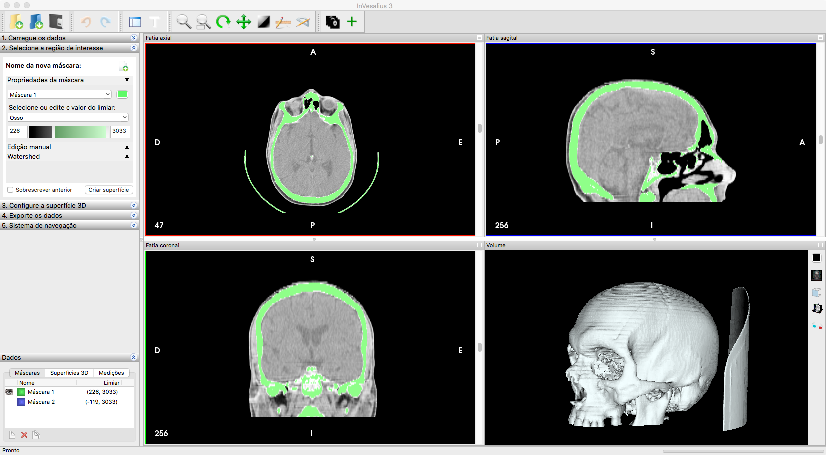
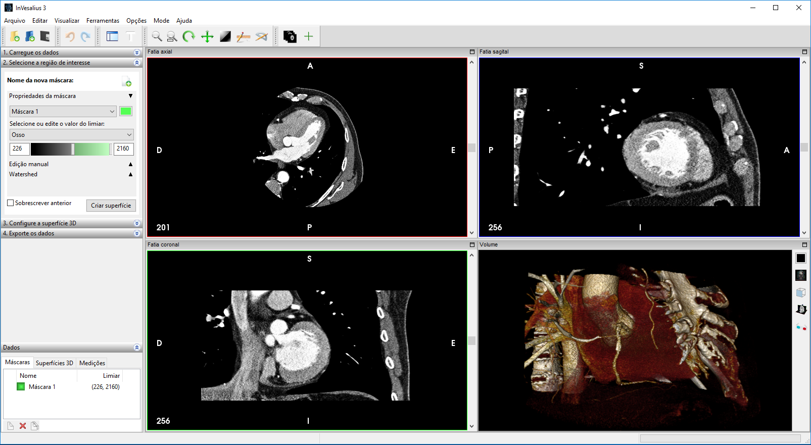
Navigating the complex world of medical diagnostics requires tools that are both highly precise and remarkably intuitive. InVesalius 3D medical imaging reconstruction software bridges the gap between raw diagnostic data and actionable anatomical insight. Developed by the Centro de Tecnologia da Informação Renato Archer, this open-source application excels at translating standard, sequential two-dimensional CT or MRI scans into highly detailed three-dimensional virtual models.
When you first load a sequence of DICOM files into the software, the interface guides you through a logical workflow. Instead of getting lost in clinical complexity, users are presented with a clean workspace where they can slice, segment, and render complex anatomical structures with high fidelity. It provides an indispensable asset for medical education, surgical planning, and deep-dive anatomical research.
Key Features of InVesalius 3D Medical Imaging Reconstruction
The software is packed with robust tools designed to handle demanding medical visualization tasks. Here is a breakdown of what makes this platform stand out:
- Broad DICOM Compatibility: Supports ACR-NEMA versions 1 and 2, alongside DICOM version 3.0 (including various lossy, lossless JPEG, and RLE encodings).
- Advanced Segmentation Options: Utilize watershed-based segmentation, region growing, or pre-defined threshold ranges tailored to specific tissue types.
- Interactive Editing Tools: Refine your models directly on 2D slices using intuitive brush-style editing features.
- High-Quality Surface Generation: Export your finalized 3D surfaces into industry-standard formats such as binary STL, OBJ, VRML, and Inventor.
- Sophisticated Volume Rendering: Features high-quality volume rendering projections, crop planes, and helpful visualization modes like Minimum, Maximum, or Mean Intensity Projection.
Who is this Medical Software For?
InVesalius is primarily designed for medical educators, researchers, and clinicians who need to visualize anatomical structures beyond flat 2D slices. Because it runs seamlessly on Linux, it is a highly accessible option for universities, research labs, and healthcare institutions globally. Its extensive internationalization—supporting languages from English and Spanish to Japanese and German—ensures that medical teams worldwide can collaborate using a shared technical framework.
If you are looking to explore this powerful diagnostic and educational aid, you can find more information and access the application directly on its official Flathub store page.
Rate the App
User Reviews












Popular Apps










Editor's Choice